
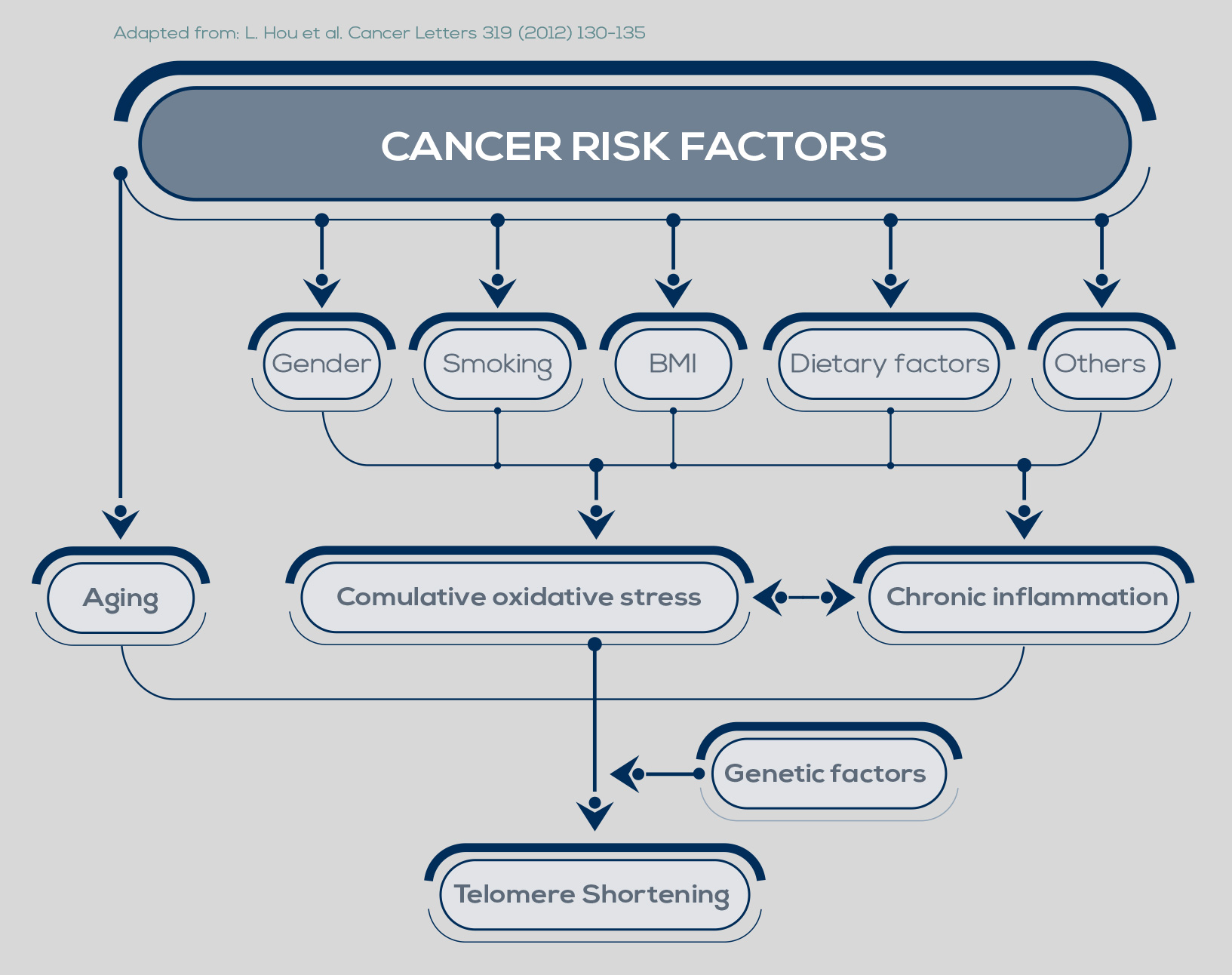
Telomere shortening is a natural phenomenon that occurs with aging. Its cause lies in the mechanism of DNA replication. In fact, one of the two strands of the DNA double helix cannot be copied all the way to its very end, leading to the loss of part of the protective cap of the chromosome at every cell cycle (50- 200 bp, depending on the tissue). After 50 to 60 cell divisions, telomeres are short enough to uncap the chromosome end, and their shortening can promote genome instability.
Inflammation and oxidative stress, two major pathways of carcinogenesis, accelerate telomere shortening. Moreover, the progressive reduction of telomere length is particularly dangerous when combined with changes such as loss of the function of p53, a protein that inhibits cell proliferation and induces senescence, or of other cell-cycle regulators.

In fact, during the period of replicative senescence (or mortality stage 1, M1) that follows the uncapping of one or two shortened telomeres, cellular proliferation is usually inhibited. However, oncogenic changes can promote an M1 bypass, thus extending the cell division period during which telomeres can further shorten. During this new dysfunctional state (the M2 crisis), chromosome end-toend fusions can occur. The cell death triggered by this situation protects from cancer development. However, between 1 in 100,000 and 1 in 10 million cells can develop mechanisms to maintain its very short telomeres, bypassing the M2 crisis and becoming immortalized.

Moreover, maintaining shortened telomeres allows the cell to express specific genes required for cancer progression that would be otherwise silenced by the conformation of telomeres.
Currently, these complex mechanisms are considered as both biomarkers and potential targets for cancer diagnosis and treatment. In 85-95% of cancers, they involve the activation of the ribonucleoprotein enzyme telomerase. Usually inactive in the vast majority of somatic cells, telomerase is able to synthesize the typical DNA repeats located at the end of chromosomes. It works as a reverse transcriptase – that is, it synthesizes DNA based on an RNA template. This RNA template is an essential component of telomerase known as human telomerase RNA (hTR) or human telomerase RNA component (hTERC).
Besides being the template for telomerase action, hTERC is involved in the catalysis, the localization, and the assembly of the enzyme too. The other essential component is the catalytic protein subunit, known as telomerase reverse transcriptase (TERT) and encoded by the hTERT gene. In the holoenzyme, TERT and hTERC interact with accessory proteins such as DKC1, NOP10, NHP2, pontin/reptin, and GAR1.
hTERC is constitutively expressed in somatic cells, whereas hTERT is epigenetically silenced, representing a limiting factor. The consequent repression of telomerase activity is a tumor suppression pathway that limits cancer cell development.

Evidence in support of this tumor suppression activity includes the association between longer telomere length and increased B cell lymphoma and chronic lymphocytic leukemia risk, as well as the association between genetic determinants of long telomeres and increased overall risk of cancer (especially melanoma and lung cancer). Acquisition of telomerase activity by cancer cells is achieved by reexpression of hTERT. Promoter mutations are present in multiple cancer types (e.g. melanoma, pleomorphic dermal sarcoma, myxoid liposarcoma, glioma, urothelial cell carcinoma, carcinoma of the skin, liver cancer, gastric cancer, pancreatic cancer, nonsmall cell lung cancer, gastrointestinal stromal tumors) and have been detected across all stages and grades of the disease, suggesting they are an early event in the process of cancer development. However, they are not indispensable: in cancer cells without these mutations, telomerase activity maintains the telomeres, too. In some cases, telomerase activity is further enhanced by other gene mutations. Beside promoter mutations, other mechanisms control telomerase expression (e.g. genomic rearrangements, alternative splicing, DNA methylation, histone modification).

Finally, in some cell types hTERC expression is a limiting factor, and the expression of both hTERT and hTERC is needed for a robust telomerase activity. The telomere elongating ability of cancer cells does not necessarily correspond to longer telomeres. Indeed, in a 2017 study that analyzed telomere length across 31 types of cancer, shorter telomeres were present in 70% of them. In fact, telomerase preferentially elongates the shortest telomeres. Available data suggest that immediately after the acquisition of hTERT promoter mutations, telomerase activation is not sufficient to elongate all telomeres. Thus, the enzyme activity concentrates on the shortest telomeres, which are elongated; meanwhile, longer telomeres shorten, resulting in gradual attrition of all the telomeres in the cell. After telomeres have shortened, the activity of the enzyme would increase to levels that are sufficient to stabilize their length.

It is also possible that during the development of cancer various cell populations arise, with long to short telomeres, and that only the cells with shortest telomeres increase telomerase activity to maintain them and avoid crisis. Furthermore, shelterin proteins may play a role by stabilizing shorter telomeres. Compared to the noncancerous cells, many cancer cells are characterized by elevated shelterin levels, and their expression correlates with the progression level of the tumor. Unfortunately, cancers with shorter telomeres are characterized by a poor prognosis. Moreover, alteration of telomere length seems to affect the morphology of cancer tissue and the expression of cancer-associated genes.

Thus, besides avoiding the M2 crisis, shortened telomeres might contribute to the severity of the tumor by influencing its genetic signature. Telomere length maintenance is not the only way telomerase participates in cancer development. In fact, this enzyme is involved in other activities that are thought to significantly contribute to oncogenesis (gene expression regulation, cell proliferation, apoptosis, WNT/β-catenin signaling, NF-kB signaling, MYC-driven oncogenesis, DNA damage repair, cell adhesion and migration, epithelial–mesenchymal transition). Because of its expression in the majority of cancer types and in cancer stem or stem-like cells, and because of its low activity in normal cells (including stem cells), anti-telomerase therapeutics could selectively induce cell death in cancer cells, sparing or minimizing the effect on noncancerous cells.

Molecules such as competitive inhibitors of telomerase or immunotherapeutic drugs were developed for the treatment of various cancers. Also, telomerase-mediated telomere-disrupting approaches, such as the use of modified nucleosides, which are incorporated into telomeric DNA by telomerase leading to telomere dysfunction, may provide a valuable option for cancer treatment. It should be noted that a limited number of cancer cells (5-15%) develop mechanisms other than telomerase activation to maintain shortened telomeres. These mechanisms are referred to as Alternative Lengthening of Telomeres (ALT) and involve the use of DNA recombination to extend telomeres length. For example, 20 to 65% of sarcomas elongate telomeres by activating ALT.