For many years, inflammation has been referred to as the response of the organism to tissue injury and infection. However, lots of things have changed since Aulus Cornelius Celsus (ca 25 BC – ca 50 AD) first defined acute inflammation as a state characterized by the presence of swelling, pain, redness and warmth – hence the name, derived from “flame”.
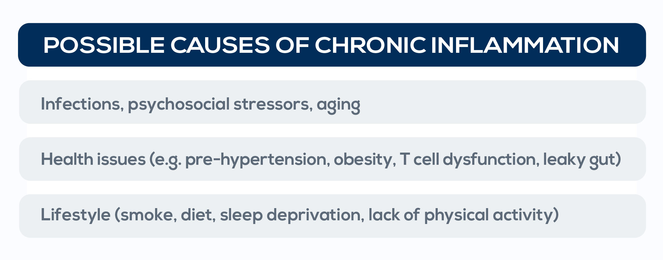
In the 19th century the father of modern pathology, Rudolph Virchow, concluded that there were various inflammatory processes. Today, it is known that inflammation can be induced by tissue stress and malfunctioning while in the absence of infections or overt tissue damage. In particular, low-grade chronic inflammation is triggered by sentinel immune cells that monitor for tissue stress and malfunctioning. It has also been associated with a number of lifestyle factors (such as smoke, unhealthy diets, sleep deprivation, and low levels of physical activity) and new correlations are constantly emerging.
Among the health issues associated with low-grade chronic inflammation is cancer. Years of research led to the conclusion that immunity serves both as a tumor suppressor and as an initiator or promoter of the disease, and that chronic inflammation is among the factors responsible for these associations. Moreover, chronic inflammation causes genomic instability. Therefore, evidence of genomic instability provokes the need to analyze the presence of inflammation as both a possible cause of the observed instability and a cancer-promoting factor. It is currently recognized that inflammation is present when the concentration or the activity of elements involved in innate responses, such as pro-inflammatory cytokines, is increased. CYTOBALANCE is the inflammation monitoring program that makes finding out and correcting the increase of these molecules possible.
Through the periodic monitoring of blood cytokine levels in apparently healthy people, CYTOBALANCE allows for intercepting an important cancer driver well before disease development and taking action against it.
Different individuals develop low-grade chronic inflammation in different ways. The inflammatory tone can increase progressively during several years or decades, depending on genetics, anatomical features, immunological history (early critical immunological experiences – such as intrauterine stimuli, type of birth, neonatal feeding, and the early use of antibiotics – and events in adulthood – such as infections), and lifelong lifestyle habits (e.g. diet). Macromolecular damage, metabolism, epigenetics, stress, proteostasis, and stem cell regeneration are all interconnected factors influencing this peculiar state of inflammation. Their effect becomes more and more evident with aging, when the chronic activation of the innate immune system can become damaging, giving rise to a form of inflammation occurring in the absence of infection that is called “inflammaging”.
Inflammaging is primarily driven by endogenous signals, including the presence of cell debris, misplaced selfmolecules, and misfolded or oxidized proteins. There are several cellular and molecular mechanisms involved: cellular senescence; the dysfunction of mitochondria (the powerhouses of the cell); defective mechanisms of autophagy and mitochondria degradation; the activation of the intracellular multiprotein complex that detects biological and non-biological stressors (the inflammasome); the dysregulation of the system controlling protein degradation; the activation of the DNA damage response; changes in the composition of the microbiota (dysbiosis); and nutrient excess and overnutrition, which can generate a specific type of chronic inflammation called “metaflammation” associated with metabolic diseases such as obesity and type 2 diabetes. All these stimuli converge on a small number of sensors which trigger the innate immune response causing inflammation and an adaptive metabolic response. This process is critical for survival until middle age, but with aging the inflammatory response usually increases, becoming detrimental – and eventually leading to chronic inflammation – in the post-reproductive age. The accumulation of senescent cells, the hyperactivation of immune response, and, according to the garbage accumulation theory, the age-related progressive impairment of cell debris elimination systems largely sustains this phenomenon. Metaflammation contributes to the onset of insulin resistance, activating inflammatory responses that affect organs such as liver and adipose tissue. This situation leads to an increased risk for metabolic syndrome, which, in turn, is associated with increased cancer risk. Lipids play a central role in this process, increasing oxidative stress and cytokines such as IL-6.

After a high-fat meal, blood levels of lipopolysaccharide (LPS, the endotoxin associated with sepsis) increase too; this generates a state called metabolic endotoxemia associated with low-grade inflammation. Finally, high-fat diets can alter gut microbiota, further increasing LPS production. Dysregulation in meal timing contributes to these metabolic and inflammatory alterations, whereas nutrient-dense diets induce the increase of adipocyte size, until they reach a structurally critical condition that contributes to metaflammation. Another inflammation-causing agent is mitochondrial DNA (mtDNA) that is released from mitochondria. Mitochondria evolutionarily derive from energy-producing bacteria which have been engulfed by ancestral cells approximately 2 billion years ago. That THE CAUSES OF INFLAMMATION is why its constituents (including freely circulating mtDNA) are immunogenic in a way similar to bacterial molecules – and that is why when released into the cytosol and extracellular environment they trigger innate immune responses, promoting inflammation.
In particular, the pro-inflammatory action of mtDNA depends on its oxidation. The highly oxidative extracellular environment typical of chronic inflammatory diseases may overwhelm antioxidant systems, further enhancing its pro-inflammatory potential. Plasma mtDNA levels are strongly variable between individuals. Almost 20% of this variability is explained by a familial component. Other factors potentially influencing it are mutations or drugs affecting intracellular mtDNA copy number, immune mechanisms controlling the response to inflammatory triggers, and the propensity to cell death, which can be enhanced in response to common chronic viral infections, such as cytomegalovirus infections in southern European populations. Moreover, mtDNA plasma levels gradually increase after the fifth decade of life. This association is not a mere epiphenomenon: the increase in plasma mtDNA can contribute to the onset and maintenance of the pro-inflammatory status typical of inflammaging. Higher mtDNA plasma levels correlate with higher amounts of proinflammatory cytokines, in particular Tumor Necrosis Factor TNF-α and IL-6. Also, blood mtDNA is significantly higher in a number of cancers, including ovarian, testicular, prostate, bladder, renal cell and lung cancer.

